Detached Retina
The retina forms the inner lining at the back of the eyeball. The vitreous is the fluid which fills the eyeball between the retina at the back and the iris and pupil at the front. A retinal detachment is caused by the vitreous fluid passing through a hole or tear in the retina, causing the retina to balloon away from the its’ position at the back of the eye. A detached retina does not provide vision, until repair is achieved.
A person's retina works like the film in a camera — it collects all the images received by the eye, instantly "develops" them and simultaneously transmits those images to the brain.
The retina is the inner layer of the eye ball, and may sometimes get split from the outer layers, and become detached.
Detached retinas are more likely to occur after middle age and affect people who are near-sighted.
How does the retina become detached?
Tiny tears or holes in the retina are usually caused by aging of the vitreous body.
The eye largely maintains its round shape from the gel-like vitreous that fills its interior and is adherent to the retina. Aging causes the vitreous to liquify and shrink away from the back of the eye. This is called a vitreous detachment and is seen very commonly after the age of 45. If an area of vitreous is quite adherent to the retina, it may cause a tear in the retina as it pulls away. Once a tear is present, there is a high risk for the vitreous fluid to seep through and cause a retina detachment.
Some symptoms
While it is normal to see "floaters" in the eye, if there is a sudden increase in the floaters or development of flashes or cobwebs, this is commonly an indication of a vitreous detachment. This process is most common in middle age when the vitreous undergoes these changes. A retinal examination is important to rule out a retinal tear or beginning of a retinal detachment.
When a retinal detachment occurs, significant peripheral and central vision loss occur, depending on the extent of the detachment. Immediate help should be sought.
Retinal tears are easily treated with laser as an office procedure. Retinal detachment is more serious and generally requires surgical repair.
Treatment
For some new or small tears, before a detachment has started, the treatment will involve placing laser around the tear to create a scar and seal the retina. However, if a retinal detachment has occurred, all the holes in the retina must be sealed and fluid accumulated beneath the retina must be drained.
Extensive detachment requires major eye surgery, usually in the Royal Alexandra Hospital.
Among the procedures and appliances that might be used in treatment are:
Lasers: sealing tears and holes by high energy light beams;
Silicone surgical explants: the sewing of flexible strips to the outside surface of the eye to compress the eyeball over tears and detached areas;
Drainage: surgically draining the fluid under the detached retina, to allow it to settle back into its regular position;
Vitreous surgery: removing the vitreous gel, hemorrhage, and membranes from the inside of the eye and from the retinal surface; and
Intraocular gas: injection of bubbles of air or special gasses into the eye to push or hold the retina in place;
Intraocular silicone oil: to help stabilize the retina in position until it seals in place.
Prognosis
More than 90% of detachments can be successfully re-attached with one surgical procedure. Should the central part of the retina — the macula — become detached for more than a few days, there is less chance of a complete return of central vision.
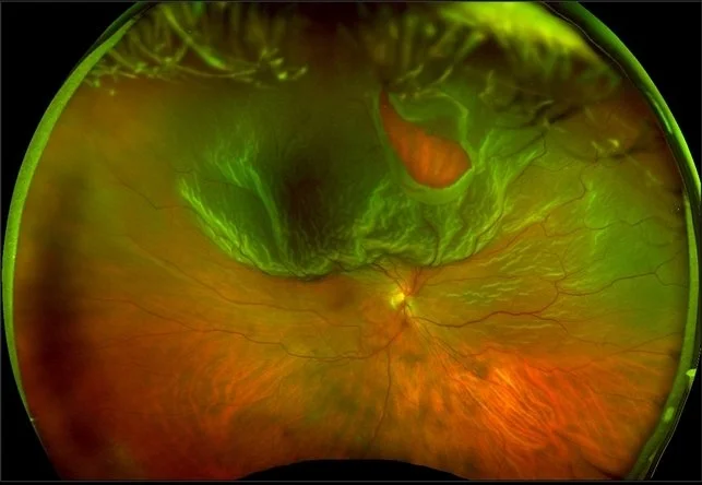
Retinal Detachment with tear in upper region of the retina.
The ribbon-like structure in motion in the video above is the retinal detachment