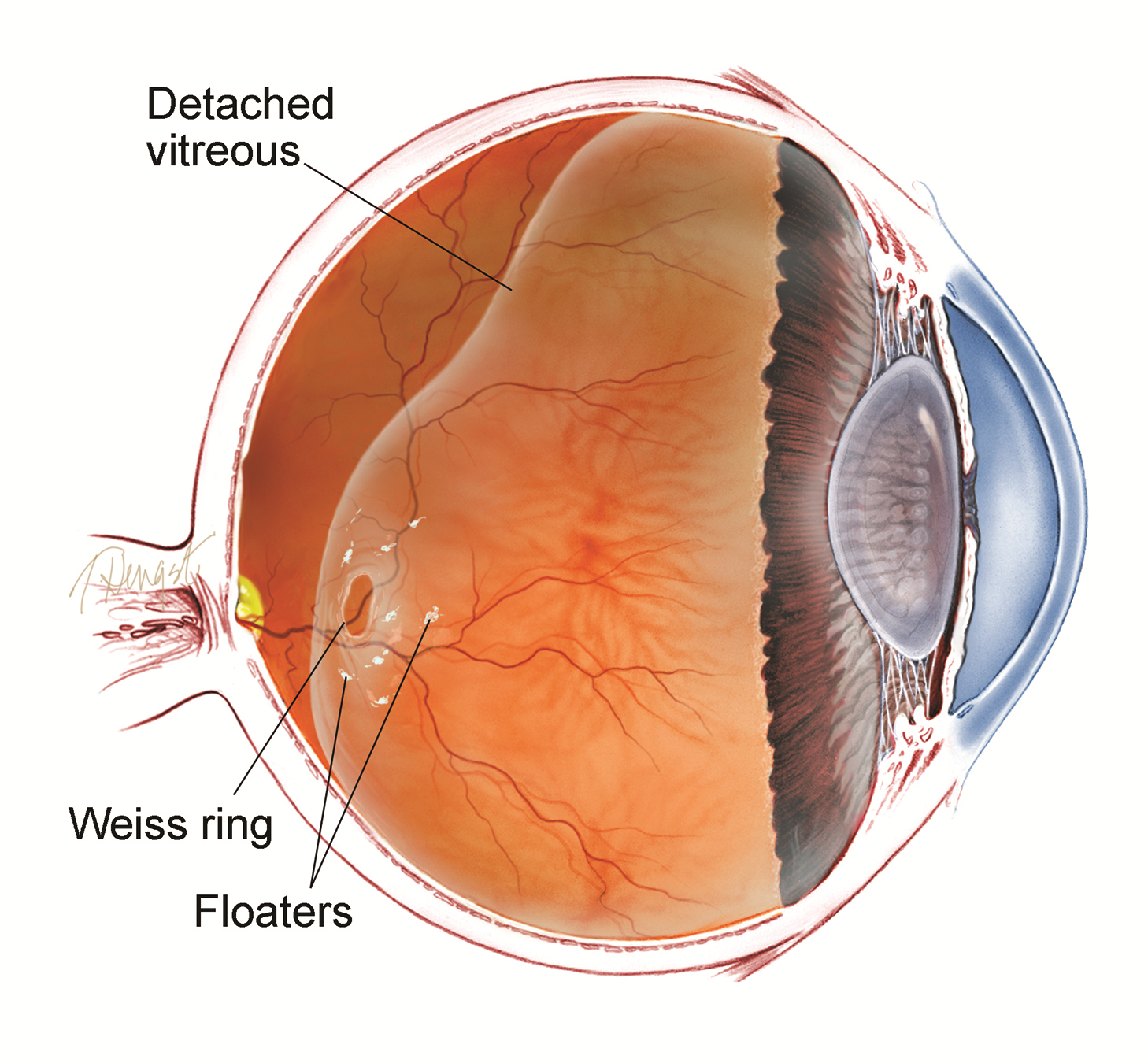
Posterior Vitreous Detachment and Floaters
The vitreous is a normally clear gel that fills most of the eyeball. With aging, the gel often develops small opacities that float around with head or eye movement casting shadows on the retina. This creates the sensation of bugs in the vision.
Patients can see these “floaters” especially against a bright background like a blue sky or a white wall. Over a period of several weeks the symptoms generally decrease and even disappear.
However, the sudden appearance of new floaters, especially if accompanied by apparent flashes of light in the peripheral vision could be a sign of a retinal tear. In this circumstance a retinal evaluation is important to rule out a tear.
When more extensive floaters develop and do not subside, this may be an indication of an intraocular inflammation or hemorrhage, a definite indication for ocular examination.
RISK FACTORS
There are many risk factors for vitreous floaters, including:
Nearsightedness
Retinal tear
Retinal detachment
Intraocular inflammation
Vitreous hemorrhage
Trauma
Previous cataract surgery can increase the perception of floaters.
Diagnostic testing
Vitreous opacities—the cause of symptomatic floaters— are detected by clinical examination with pupil dilation. This is the most valuable and reliable way to observe floaters that a patient is seeing.
Other ways to evaluate floaters include optical coherence tomography (OCT), B-scan ultrasound, and retinal photography.
Treatment
Any management of floaters will depend on the initial cause and will be discussed with the patient at the time of the examination.