Vascular Occlusions
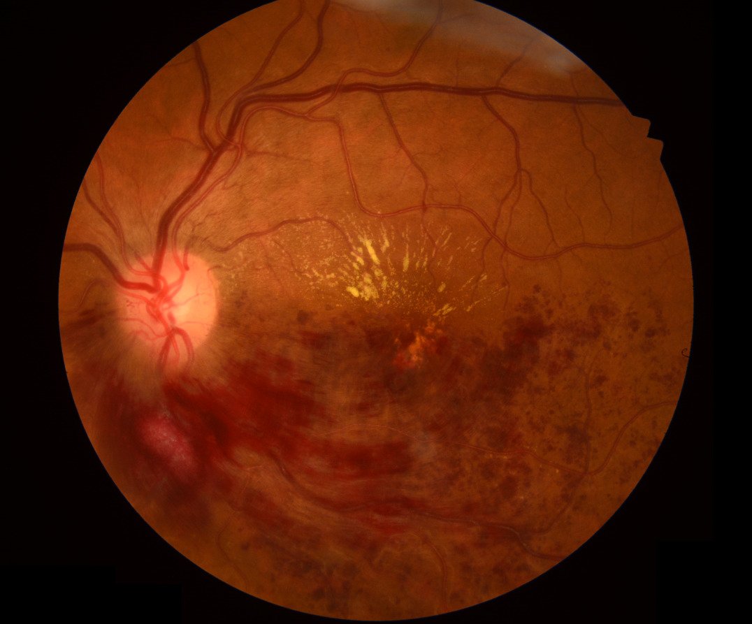
A retinal vein occlusion (vascular occlusion) occurs when a blockage in the vein prevents adequate blood flowing into the affected area of the eye. There are two types : a central retinal vein occlusion (CRVO) or a branch retinal vein occlusion (BRVO).
Understandably, the central form, which affects the main retinal vein, is the more serious, while the branch form relates to a vein blockage in one specific part of the eye.
These problems are more likely to occur among people who already suffer from:
Glaucoma
Diabetes
Age-related blood vessel problems
High blood pressure
Blood disorders
The symptoms of retinal vein occlusion include blurred vision, floaters and, at times, pain in the eye. The blurred vision occurs when blood, leaking from the blocked vein, collects in the macula — the centre of the retina. As described in the floaters section of the website, floaters can appear as spots or other shapes, interfering with normal vision. These are usually caused by the bleeding into the vitreous, causing shadows to be cast on the retina. When pain occurs, it is usually related to excessive eye pressure called neovascular glaucoma. It is a complication created when the central retinal vein is involved.
Treatment for both types is recommended when there is an accumulation of fluid into the macula (edema) and vision deteriorates. Treatment for this problem involves injectable medication in the family of anti-VEGF, similar to that used in diabetic macular edema and macular degeneration.
Laser is applied only when new fragile blood vessels grow on the surface of the retina. Should these vessels bleed, a surgical procedure is often required.
Branch retinal vein occlusion